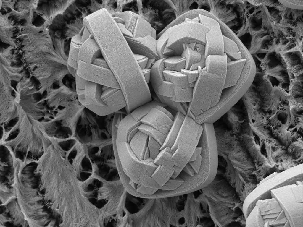
Scanning electron micrograph of three defects on the surface of a heart stent. The defects result from the crystallization of titanium dioxide out of solution during the fabrication process. Needless to say, such defects on the surface of a heart stent are undesirable. Specimen courtesy of Harald Nuhn (Alfred Mann Institute). Image credit: Clive Hayzelden.
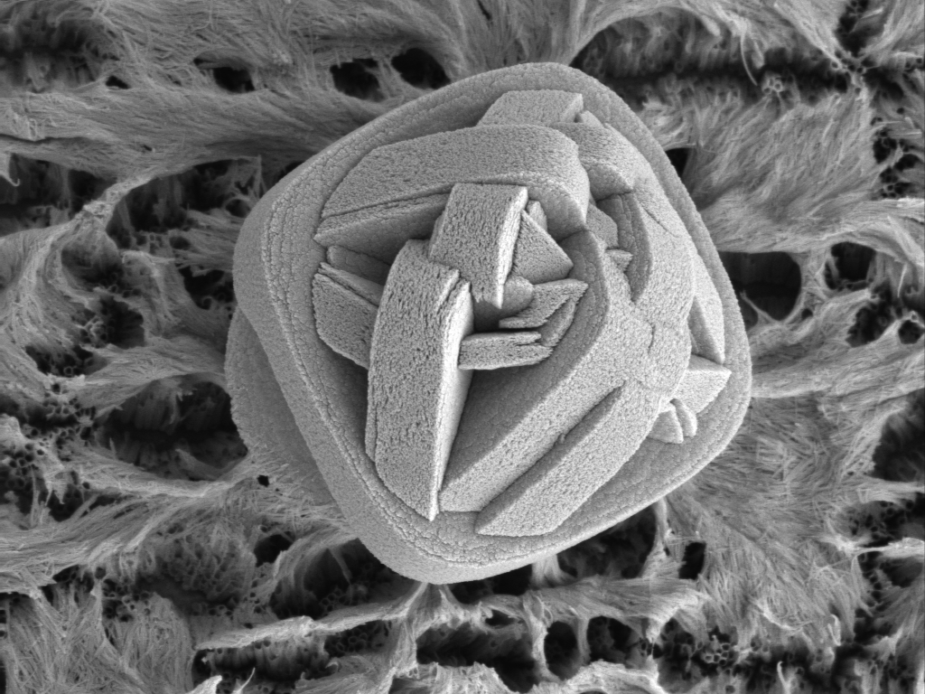
Higher magnification image of a single TiO2 defect on the surface of a heart stent. Titanium dioxide nanotubes are visible on the stent. These hollow TiO2 nanotubes are filled with slow-release drugs to limit the rejection of the stent by the human body. Specimen courtesy of Harald Nuhn (Alfred Mann Institute). Image credit: Clive Hayzelden.
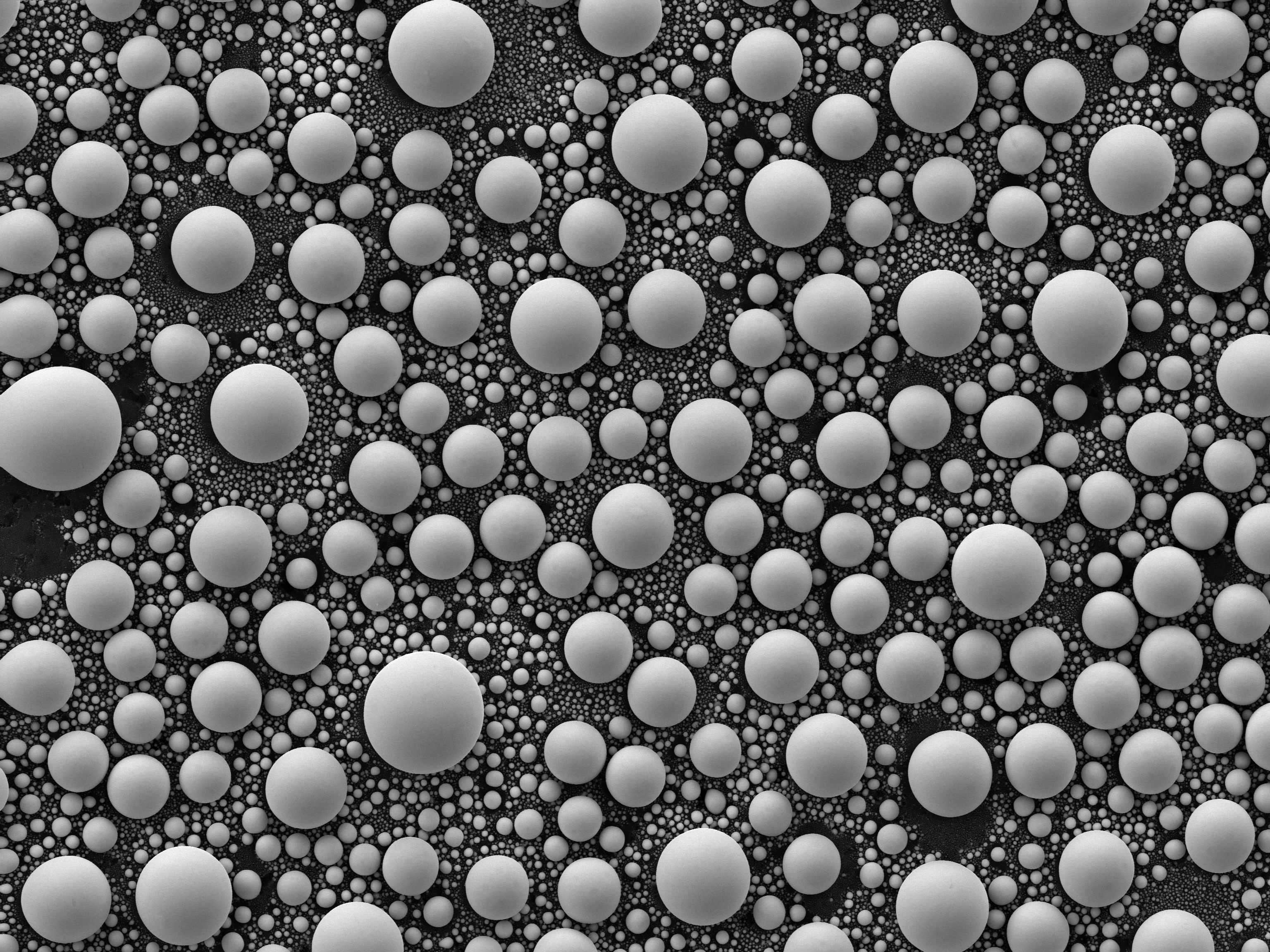
Tin balls are a widely-used imaging standard in FE-SEM, being useful to assess image quality and as an aid in correcting astigmatism for new users. When a thin-layer of tin on vitrious carbon is heated to the melting point, surface tension causes the tin to form a sphere so that the surface area / volume ratio is minimized. Image credit: Clive Hayzelden.